 Written by Hirotsugu Suwanai, MD, PhD
Journal Club of the Suwanai · Metabolism & Immunity
After a meal, blood glucose rises, insulin is secreted, and the liver stops producing glucose. What if immune cells were an essential part of this textbook sequence?
In 2020, Molecular Cell published a study showing that macrophages in adipose tissue help regulate post-meal glucose. The work came from the group of Takashi Kadowaki and Kohjiro Ueki, and I took part as a co-author. Read the original paper ↗
Key point: when an “inflammatory” signal helps metabolism ↓
Adipose macrophages make IL-10 after feeding
Macrophages are immune cells that clear pathogens and drive inflammation. In obesity, they accumulate in fat tissue and promote insulin resistance.
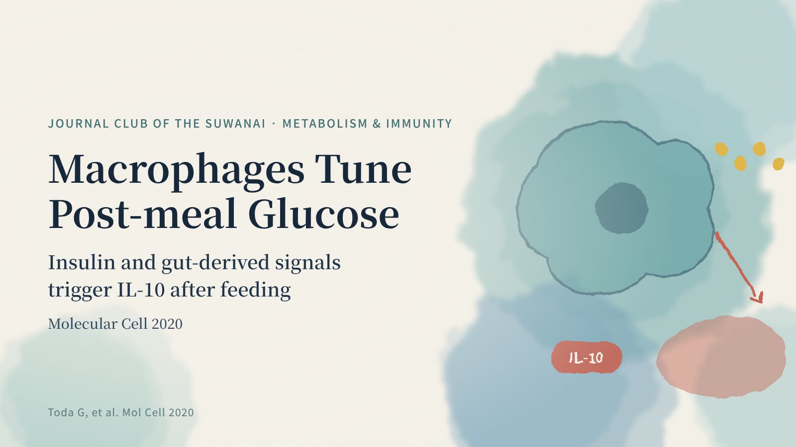
The team found that feeding prompts adipose tissue macrophages to produce interleukin-10 (IL-10), a classic anti-inflammatory cytokine. Working together with insulin, this IL-10 suppressed glucose production (gluconeogenesis) in the liver.
Two signals are required
IL-10 production depended on two signals that both rise with feeding: insulin and lipopolysaccharide (LPS) derived from the gut microbiome. Inside macrophages, these signals converged on the Akt/mTOR pathway.
- Mice lacking the insulin receptor in myeloid cells, or transplanted with bone marrow carrying mutant TLR4 (the LPS receptor), showed higher expression of hepatic gluconeogenic genes and impaired glucose tolerance.
- Myeloid deletion of Akt1 and Akt2 produced similar defects, which were rescued by additionally deleting TSC2, an inhibitor of mTOR.
The mechanism fails in obesity
In obesity, macrophages themselves become insulin resistant and post-meal IL-10 production falls. Restoring IL-10 with an adenoviral vector improved postprandial hyperglycemia.
Journal Club view: when an “inflammatory” signal helps metabolism
LPS is usually cast as a villain that worsens inflammation and insulin resistance. This study suggests that physiological, meal-related LPS together with insulin triggers anti-inflammatory IL-10 that supports glucose control. The appeal of the work lies in showing hormones, the gut environment and immune cells cooperating within the short window after a meal.
The experiments were done mainly in mice, so how much this pathway contributes to post-meal glucose in humans remains to be tested. How differences in the microbiome or diet shape this mechanism is another open question.
A new angle on post-meal hyperglycemia
Post-meal hyperglycemia appears early in type 2 diabetes. Viewing it through immune cells and the gut environment — not only insulin secretion and action — may open new therapeutic ideas.
Related articles
- MEK/ERK Signaling Supports Both the Number and the Function of β-Cells: A Study in Diabetes
- Semaglutide Extends Lifespan in Aged Female Mice: What a Nature Study Means for Diabetes Drugs and Aging Research
REFERENCE
Toda G, Soeda K, Okazaki Y, Kobayashi N, Masuda Y, Arakawa N, Suwanai H, Masamoto Y, Izumida Y, Kamei N, Sasako T, Suzuki R, Kubota T, Kubota N, Kurokawa M, Tobe K, Noda T, Honda K, Accili D, Yamauchi T, Kadowaki T, Ueki K. Insulin- and Lipopolysaccharide-Mediated Signaling in Adipose Tissue Macrophages Regulates Postprandial Glycemia through Akt-mTOR Activation. Mol Cell. 2020;79(1):43–53.e4.
doi:10.1016/j.molcel.2020.04.033 ↗
This article is an independent English summary and commentary based on the original paper. No figures are reproduced.