 Written by Hirotsugu Suwanai, MD, PhD
Journal Club of the Suwanai · Pancreatic Beta Cells
In type 2 diabetes, two problems occur together: too few insulin-producing β-cells and weaker function in each β-cell. Is there a signal that links the two?
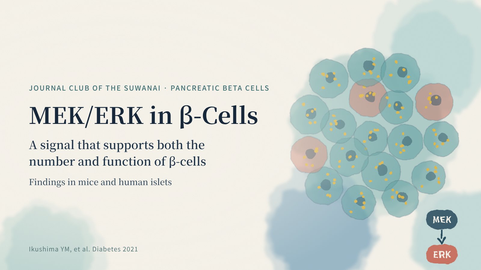
In 2021, Diabetes, the journal of the American Diabetes Association, published a study showing that MEK/ERK signaling in β-cells regulates both β-cell mass and insulin secretion. The work came from the group of Kohjiro Ueki and colleagues, and I took part as a co-author. Read the original paper ↗
Key point: how to read the “two roles” ↓
The “number” and “function” of β-cells
Insulin is secreted by β-cells in the pancreatic islets. High glucose in diabetes reflects both a β-cell mass too small for the body’s insulin needs and individual β-cells that can no longer release enough insulin in response to glucose.
The MEK/ERK pathway is a classic signaling route for cell proliferation and differentiation: MEK1/2 phosphorylate and activate ERK1/2, triggering many cellular responses. This study asked what the pathway does in β-cells, in mice and in human islets.
ERK activity is reduced in diabetic islets
The team found that active, phosphorylated ERK1/2 was significantly reduced in islets from db/db mice, a model of obesity and diabetes, and in islets from people with type 2 diabetes — an important observation in humans as well as mice. Abstract
What happens when MEK is deleted in β-cells?
Mice lacking Mek1 and Mek2 specifically in β-cells, and therefore lacking ERK signaling, developed worse glucose intolerance on a high-fat diet. Two distinct changes explained this.
Changes in β-cell-specific Mek1/2 knockout mice on a high-fat diet. Source: Ikushima et al., Diabetes (2021).
1. Less β-cell proliferation and mass
Without MEK/ERK signaling, β-cell proliferation fell and β-cell mass decreased: the mice could not expand their β-cells to meet the higher insulin demand of a high-fat diet.
2. Less secretion from each β-cell
Individual β-cells showed fewer insulin exocytosis events, and molecules involved in exocytosis were less phosphorylated — ERK signaling also governs the act of releasing insulin.
Journal Club view: how to read the “two roles”
β-cell growth and insulin secretion are often studied as separate mechanisms. This study’s significance lies in showing, with genetically engineered mice, that one signaling pathway spans both. Nature Reviews Endocrinology highlighted the work from the perspective of the temporal control of ERK signaling in β-cells. Related commentary ↗
The authors suggest the pathway could inspire new diabetes drugs. Yet MEK/ERK drives proliferation in many cells throughout the body, and cancer therapy uses drugs that inhibit it. Harnessing it in β-cells would require selectivity and the right strength and timing.
Reduced ERK activity in human islets is an association; this observation alone cannot tell whether it causes diabetes or results from hyperglycemia. How far the mouse findings apply to human β-cells, and at what stage of disease the pathway matters most, are open questions.
Where this fits in metabolic research
Protecting β-cell mass and function is central to slowing diabetes. Hormones such as GLP-1, which stimulate insulin secretion, are also known to activate ERK in β-cells. Viewing β-cell “growth” and “work” through one pathway may also help us understand how existing drugs act.
REFERENCE
Ikushima YM, Awazawa M, Kobayashi N, Osonoi S, Takemiya S, Kobayashi H, Suwanai H, Morimoto Y, Soeda K, Adachi J, Muratani M, Charron J, Mizukami H, Takahashi N, Ueki K. MEK/ERK Signaling in β-Cells Bifunctionally Regulates β-Cell Mass and Glucose-Stimulated Insulin Secretion Response to Maintain Glucose Homeostasis. Diabetes. 2021;70(7):1519–1535.
doi:10.2337/db20-1295 ↗
This article is an independent English summary and commentary based on the abstract of the original paper. No figures are reproduced.